Coracohumeral Ligament
Introduction
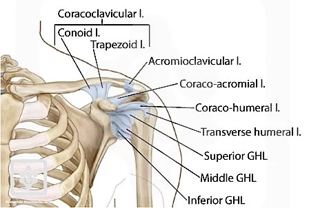
The coracohumeral ligament is crucial in the shoulder joint. The larger tubercle of the humerus and the coracoid process of the scapula are joined by this ligament. Restricting excessive external rotation and humeral abduction, helping to stabilize the shoulder joint.
It also helps to prevent dislocation and distribute pressures throughout the joint. The glenohumeral and coracohumeral ligaments cooperate to give the shoulder joint stability and functionality.
Structure of the Coracohumeral Ligament
A robust and substantial band of connective tissue, the coracohumeral ligament connects the scapula’s coracoid process to the humerus’s greater tubercle. One of the main ligaments that helps in joint stability, it is situated on the anterior portion of the shoulder joint.
Dense collagen fibers aligned parallel to one another make up the ligament. These fibers provide the ligament its strength and help in its ability to withstand tensile stresses. Additionally, the ligament has elastic fibers that enable it to gradually stretch while under pressure, reducing the likelihood of injury and absorbing shock.
The superior and inferior bands are the two distinct sections that make up the coracohumeral ligament. The majority of the ligament’s stabilizing properties are provided by the superior band, which is thicker and stronger than the inferior band. Because the inferior band is thinner and more elastic, the shoulder joint can move more freely.
Together with other ligaments and muscles, the coracohumeral ligament gives the shoulder joint stability and functionality. It helps in preventing excessive humeral abduction and external rotation, which can result in injury or dislocation. Additionally, it contributes to the distribution of stresses throughout the joint, preventing harm to the surrounding tissues.
All things considered, the coracohumeral ligament is a crucial component of the shoulder joint that helps in function and stability. It is a vital part of a healthy and functional shoulder because of its robust and elastic qualities, which enable it to tolerate the forces put on the joint during movement.
Attachment of the Coracohumeral Ligament
Two distinct bone landmarks in the shoulder joint—the larger tubercle of the humerus and the coracoid process of the scapula—are where the coracohumeral ligament is attached.
A bony protrusion that protrudes anteriorly from the scapula is called the coracoid process. The base of the coracoid process, close to the origin of the short head of the biceps brachii muscle, is where the superior band of the coracohumeral ligament connects. The ligament can stabilize the shoulder joint during flexion and internal rotation of the humerus because of this attachment point.
A bony protrusion on the lateral surface of the humerus, the greater tubercle is situated somewhat above the shoulder joint. The smaller tuberosity, a tiny depression on the greater tubercle, is where the inferior band of the coracohumeral ligament is attached. The ligament can stabilize the shoulder joint during motions including external rotation and humeral abduction because of this attachment point.
When combined, these two attachment sites enable the coracohumeral ligament to stabilize the shoulder joint in a variety of motion planes. The ligament helps in limiting excessive movement and possible injury to joints by attaching to the scapula and humerus.
Function of the Coracohumeral Ligament
One significant structure that is essential to maintaining the stability of the shoulder joint is the coracohumeral ligament. The coracoid process of the scapula and the larger tubercle of the humerus are joined by this robust, fibrous band. The superior and inferior bands, two separate bands that adhere to various bony landmarks in the shoulder joint, make up the ligament.
Stability of the shoulder joint during movement is the coracohumeral ligament’s main purpose. This is especially crucial when the humerus is being moved in flexion, extension, internal and external rotation, and abduction. By acting as a restriction, the ligament helps to avoid overuse and possible joint injury.
The superior band of the coracohumeral ligament attaches to the base of the coracoid process to provide support during flexion and internal rotation of the humerus. The ligament is able to withstand severe anterior translation of the humeral head thanks to this attachment point, which would otherwise cause the joint to dislocate or subluxate.
By connecting to the humerus’s greater tubercle at the lesser tuberosity, the inferior band of the coracohumeral ligament stabilizes the humerus during external rotation and abduction. The ligament can withstand severe posterior translation of the humeral head because of its attachment site, which would otherwise cause instability or impingement in the joint.
Apart from offering support during motion, the coracohumeral ligament also contributes to protecting the stability of other shoulder joint components. For instance, it keeps the biceps tendon from slipping out of its groove in the humerus and supports its long head.
Ultimately, the coracohumeral ligament is a crucial component for keeping stability and avoiding shoulder joint injuries. It is an essential part of the intricate biomechanics of the shoulder because of its attachment locations on the scapula and humerus, which enable it to offer stability in a variety of planes of motion.
Blood supply of the Coracohumeral Ligament
There are multiple sources of blood flow to the coracohumeral ligament. The suprascapular artery, which emerges from the subclavian artery’s thyrocervical trunk, is the main source. The coracohumeral ligament is one of the branches that the suprascapular artery produces to supply the shoulder joint.
Additionally, the coracohumeral ligament receives blood from the anterior and posterior circumflex humeral arteries. These arteries supply collateral circulation to the shoulder joint and emerge from the axillary artery.
The coracohumeral ligament’s blood flow is crucial to maintaining its structural soundness and avoiding injury. Shoulder joint instability and dislocation brought on by ligament injuries may result in chronic pain and functional impairment. For the shoulder to operate at its best, enough blood supply to the ligament must be maintained.
Clinical Significance
Coracohumeral Ligament Injury
A coracohumeral ligament injury happens when trauma, repetitive overhead motions, or severe strain cause the coracohumeral ligament—a crucial structure that aids in shoulder joint stability—to become strained, partially tear, or injured.
Symptoms of the coracohumeral ligament
Depending on the severity of the injury, coracohumeral ligament symptoms can change. Typical symptoms include the following:
- Shoulder Pain: The most typical sign of an injury to the coracohumeral ligament is pain. When the arm is raised or moved, the pain might get worse.
- Weakness: Shoulder and arm weakness may result from injuries to the coracohumeral ligament. It could be difficult to raise or move the arm due to this weakness.
- Shoulder joint Instability: The shoulder joint is stabilized in large part by the coracohumeral ligament. Ligament injuries can result in joint instability, which can cause subluxation or dislocation.
- Restricted range of motion: Shoulder joint range of motion may be restricted by injuries to the coracohumeral ligament. Daily tasks like lifting objects or reaching overhead may become difficult as a result.
- Bruising and swelling: In more difficult situations, the shoulder joint may sustain bruising and swelling.
If someone experiences any of these symptoms, they should seek medical attention. After diagnosing the injury, a medical professional can suggest the best way to take action, which may involve surgery, physical therapy, or rest.
Coracohumeral Ligament Injury Causes
A tiny but crucial ligament in the shoulder joint is the coracohumeral ligament. It helps in stabilizing the shoulder joint while in motion by joining the scapula’s coracoid process to the humerus’ greater tubercle. The coracohumeral ligament can be injured by several activities, such as:
- Repeated overhead motions: The coracohumeral ligament can be severely strained by activities that require repetitive overhead motions, such as painting, swimming, or throwing a ball. This may cause a decrease over time and ultimately lead to an injury.
- Trauma: The coracohumeral ligament may sustain a sudden injury from a direct hit to the shoulder or from falling onto an outstretched arm. Contact sports and other activities where there is a high risk of falls are more likely to cause this kind of injury.
- Poor posture: Injuries to the coracohumeral ligament can also be caused by poor posture. To increase the risk of injury, slouching or hunching over can put additional strain on the ligament.
- Shoulder impingement: This condition happens when the rotator cuff tendons and other soft tissues in the shoulder get squeezed between the shoulder joint’s bones. Injuries may result from this increased strain on the coracohumeral ligament.
- Aging: Our ligaments and other connective tissues lose their elasticity and become more exposed to injuries as we age. This may raise the risk of injury and make the coracohumeral ligament more prone to worsening.
Preventing coracohumeral ligament injuries requires taking the right precautions, which include maintaining proper posture, exercising with proper technique, and gradually increasing the duration and intensity of your workouts. To stop further damage and encourage healing, get medical help if you show signs of a coracohumeral ligament injury.
Diagnosis
A substantial band of connective tissue called the coracohumeral ligament joins the scapula’s coracoid process to the humerus’s greater tubercle. It inhibits the humeral head’s poor translation and excessive external rotation while stabilizing the shoulder joint.
A thorough physical examination is required to diagnose a coracohumeral ligament injury. The actions listed below can be taken:
- Medical history: The doctor will inquire about the patient’s symptoms, such as pain, weakness, and range-of-motion restrictions.
- Physical examination: For the purpose to look for signs of weakness or instability, a doctor will physically examine the shoulder joint. To evaluate the strength of the rotator cuff muscles and other shoulder joint structures, they may use specific tests like the lift-off test or the belly-press test.
- Imaging tests: Imaging tests may be needed if the physical examination is insufficient to make a diagnosis.MRIs can give specific images of soft tissues, including ligaments, while X-rays can help rule out other disorders like arthritis or fractures.
- Diagnostic injection: In some situations, a diagnostic injection may be used to confirm a diagnosis. After injecting a local anesthetic and steroid medication into the shoulder joint, if the patient gets a significant reduction in pain, this could be a symptom of a coracohumeral ligament injury.
To diagnose a coracohumeral ligament injury, a combination of imaging investigations, physical examination, medical history, and diagnostic injections may be used. Depending on how severe the injury was, treatment options could involve surgery, physical therapy, medication, or rest.
Special test
Coracoid impingement test
A special test for determining the stability of the coracohumeral joint is the coracoid impingement test. The humerus bone in the shoulder blade socket is stabilized by this ligament, which is found in the shoulder joint.
In the coracoid impingement test, the patient is either sitting or standing with their arm by their side. After that, the examiner takes hold of the patient’s elbow and internally rotates their arm, flexing it to 90 degrees. The examiner then rests their other hand on the bony protuberance on the front of the shoulder blade called the coracoid process.
After applying pressure to the coracoid process, the examiner asks the patient if they feel any pain or discomfort. An injury to the ligament may be suggested if the maneuver causes pain or discomfort in the coracohumeral ligament area.
The coracoid impingement test is frequently used to evaluate shoulder injuries in combination with other specialized testing and physical examination methods. Due to the possibility of other shoulder joint components causing pain or discomfort during this movement, a positive test result might not always suggest a coracohumeral ligament injury.
Treatment of the Coracohumeral Ligament
Conservative treatment
The goal of conservative treatment for a coracohumeral ligament injury is to lessen shoulder joint discomfort and inflammation while improving function. Some conservative methods of treating the coracohumeral ligament include the following:
- Rest: The shoulder joint must heal. Rest and avoid activities that aggravate the pain. Immobilizing the shoulder and easing the strain on the injured ligament can be performed with a sling.
- Ice therapy: Applying ice to an injured area can help reduce inflammation, discomfort, and swelling. Applying ice should be done multiple times a day for 20 minutes at a time.
- Physical therapy: A physical therapist can create an exercise program to strengthen the muscles around the shoulder joint and improve the range of motion. Exercises may include stretching, range-of-motion exercises, and strengthening exercises.
- Medication: Ibuprofen and naproxen are examples of over-the-counter nonsteroidal anti-inflammatory medications (NSAIDs) that can help reduce pain and inflammation.
- Corticosteroid injections: If conservative measures are ineffective, corticosteroid injections may be recommended. Injections of corticosteroids can help lessen shoulder joint pain and inflammation.
- With platelet-rich plasma (PRP) therapy, a concentrated solution of the patient’s own blood platelets is injected into the injured area. Growth factors included in platelets may aid in promoting recovery.
Therefore, the basic goal of conservative treatment for a coracohumeral ligament injury is to lessen shoulder joint pain and inflammation while also restoring function. Working with a healthcare professional to create a treatment plan that is suitable for the severity of the injury is crucial.
FAQs
What is the coracohumeral ligament?
The coracohumeral ligament appears to serve as a sort of holder for the subscapularis and supraspinatus muscles, covering the whole subscapularis muscle and insertion. The ligament’s relatively high type III collagen and uneven, sparse fiber composition would indicate flexibility.
What movement does the coracohumeral ligament limit?
A coracohumeral ligament contracture limits the posterior-inferior shift of the humeral head and the external rotation of the shoulder with the arm at the side. The glenohumeral joint’s range of motion is intended to be limited by the contracture.
What is the origin of the coracohumeral ligament?
A portion of the subscapularis muscle was covered by the coracohumeral ligament, which arose from the horizontal limb and base of the coracoid process. A large portion of the anterior surface of the muscle was covered by the ligament’s superficial layer.
How is a torn coracohumeral ligament treated?
Repair or reconstruction, which involves replacing or repairing damaged ligaments using graft tissue, is a common surgery. For more complicated injuries, open surgery could occasionally be necessary. For the best healing and shoulder function, post-operative rehabilitation is crucial.
Where is the coracoacromial ligament located?
Strong and triangular, the coracoacromial ligament connects the acromion to the coracoid process. It protects the humerus’s head. During reconstructive surgery for the acromioclavicular joint (shoulder joint), its acromial attachment may be moved to the clavicle.
What is the length of the coracoacromial ligament?
Male and female coracoacromial ligaments measured 27.5 ± 2.5 mm and 27.5 ± 2.3 mm in length and 1.05 ± 0.19 mm and 0.96 ± 0.24 mm in thickness, respectively.
References:
- Contributors to Wikimedia projects. (2025). Coracohumeral ligament – Wikipedia. Wikimedia Foundation, Inc.. https://en.wikipedia.org/wiki/Coracohumeral_ligament
- Danijel Tosovic. (2024). Coracohumeral ligament. Kenhub. https://www.kenhub.com/en/library/anatomy/coracohumeral-ligament
- Dr.Ritika Patel. (2023). Coracohumeral Ligament – Anatomy, Structure, Function, Injury. SAMARPAN PHYSIOTHERAPY CLINIC AHMEDABAD. https://samarpanphysioclinic.com/coracohumeral-ligament/