Types of Muscles In the Human Body
Introduction
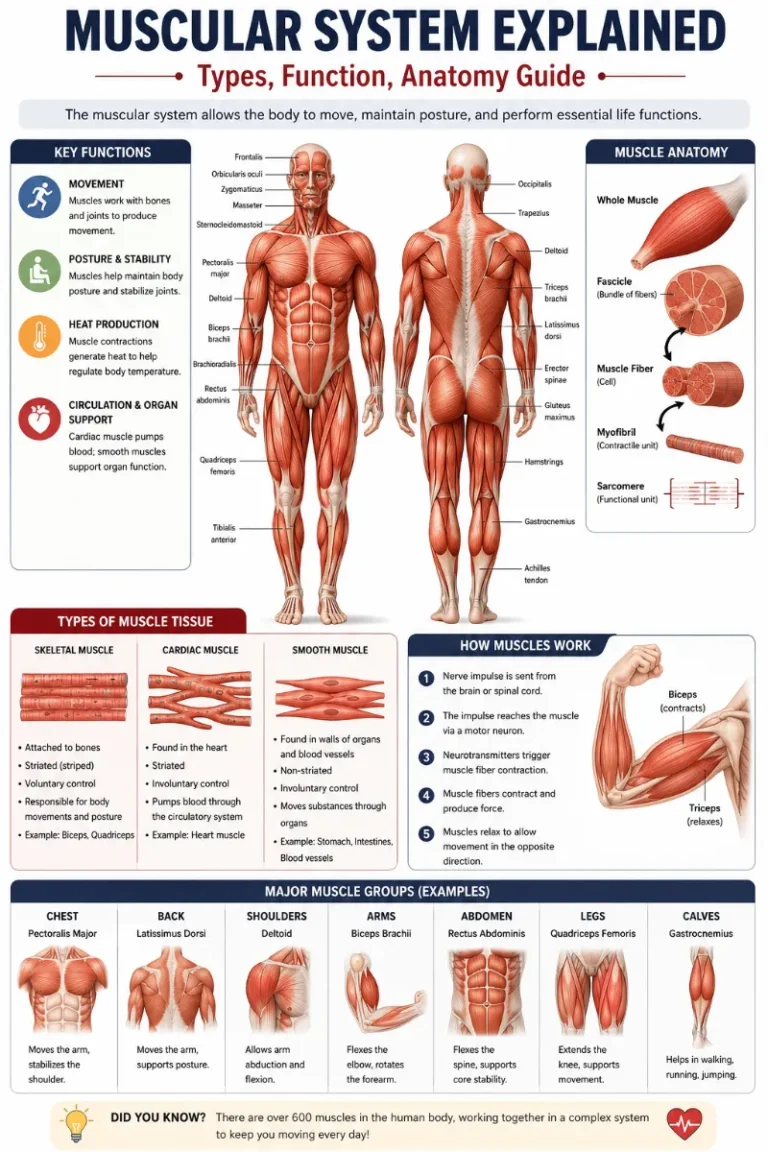
Your body’s muscles are fragments of soft tissue. They support you in everything from running a marathon to keeping your body motionless. Additionally, muscles support and move your organs. Your heart is a labor-intensive muscle that keeps you alive by beating thousands of times every day.
You have a lot of muscles; thus, diseases and injuries that affect them are common. Everybody occasionally experiences a sore muscle, but chronic muscle soreness, weakness, and other symptoms may indicate more significant problems. If you have difficulties moving or if your muscular soreness lasts more than a week, see a doctor. If breathing or swallowing becomes difficult, visit the emergency department.
Anatomy
Thousands of tiny strands are braided together to form your muscles. Your body’s organs are moved by the stretching and pressing of these fibers. Your muscles form a quilt-like structure that envelops your body. They work together to move you while you run in every direction.
Types of muscles
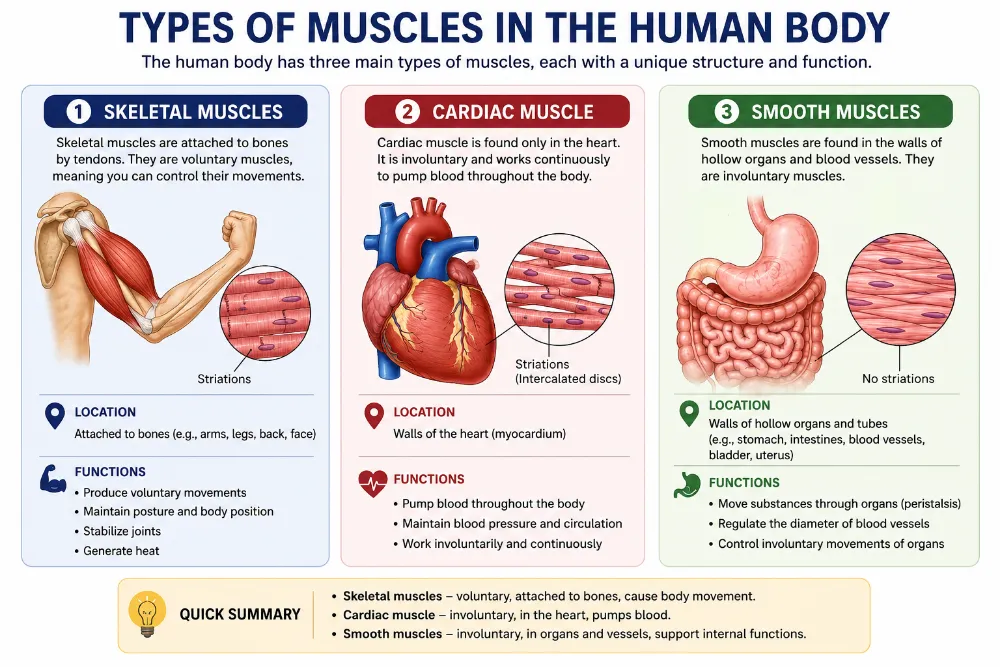
Muscles are arranged by tissue type by medical professionals. Your body contains three different kinds of muscular tissue:
- Skeletal Muscles.
- Cardiac Muscles.
- Smooth Muscles.
Skeletal Muscles
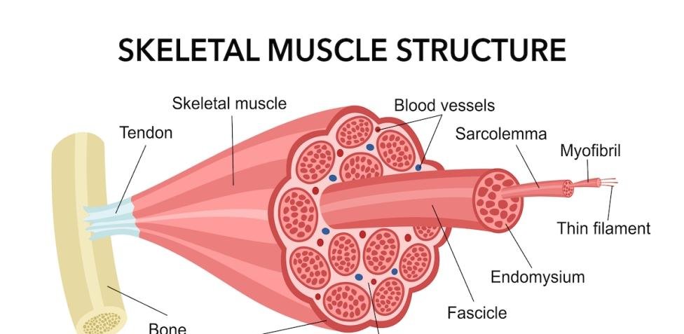
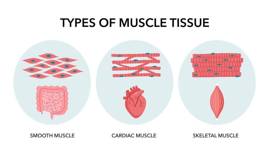
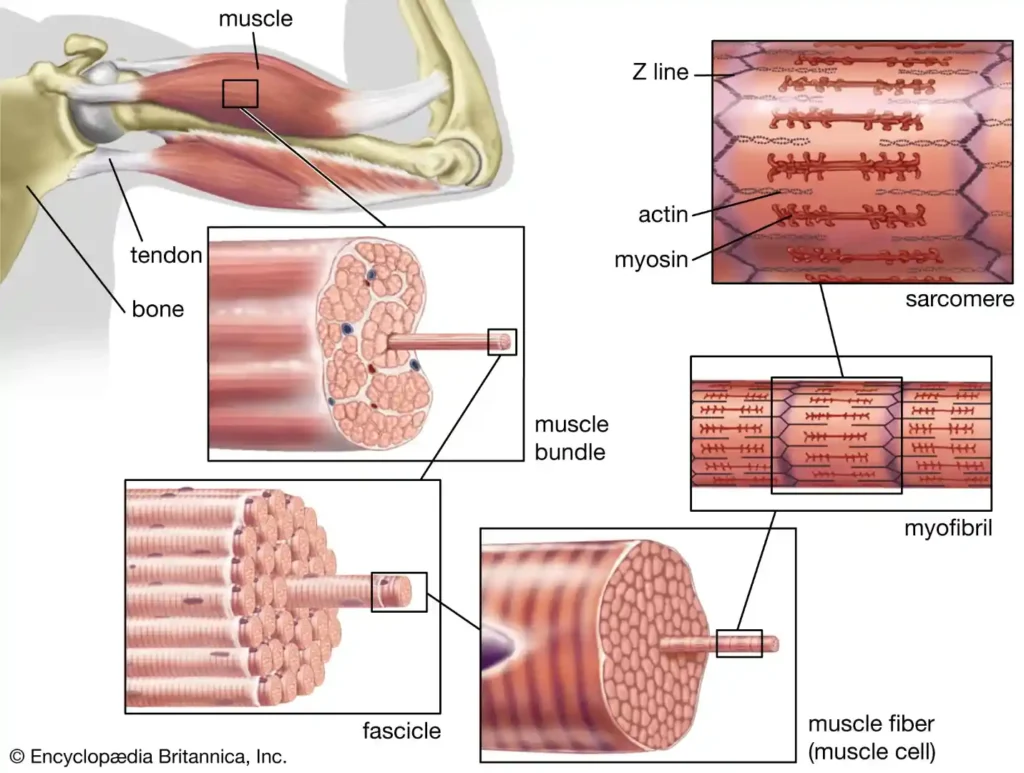
Skeletal movements are caused by skeletal muscles, which are connected to the bones. The Central Nervous System, through the peripheral nervous system (PNS), controls skeletal muscles. These muscles are therefore within cognitive, or voluntary, control. The basic unit is the multinucleated muscle fiber. Each of these striated muscle fibers functions independently of its surrounding muscle fibers and possesses transverse streaks.
Skeletal muscle is a voluntary, striated muscle that is attached to bones and is responsible for body movement. It works under conscious control and forms the largest portion of muscle tissue in the human body.
Essential Terms
- A single muscle cell is a muscular fiber.
- Fascicle: Muscle fiber bundle
- Myofibril: Inside fibers, a rod-like structure
- Sarcomere: Fundamental contraction unit
- Actin: Thin filament protein
- Myosin: Thick filament protein
- Origin: Muscle attachment that is fixed
- Insertion: Adaptable fastening
Tendon Attachment to Bone
- Strong fibrous connective tissue makes up tendons.
- They link bone and muscle.
- When the muscle contracts, the tendon pulls the bone, resulting in movement.
Muscle Interaction
Antagonistic pairs are pairs of muscles. For instance:
- Biceps (elbow flexion)
- Triceps (elbow extension)
- Smooth movement results from one muscle contracting while the other relaxes.
Anatomy of Skeletal Muscle
- Epimysium: The epimysium covers the whole muscle.
- Perimysium: Encloses fascicles
- Endomysium: Envelops individual fibers, including nerves and blood vessels.
Actin, a thin filament
- Composed of the protein actin
- Includes tropomyosin and troponin.
- Contributes to the control of muscular contraction
Myosin, or thick filament
- Composed of the protein myosin
- Has heads that attach to actin
- In charge of producing force
Blood and Nerve Supply
Supply of nerves:
- Managed by motor neurons
- The neuromuscular junction transmits the signal.
Blood supply:
- An extensive network of capillaries and arteries
- Supplies nourishment and oxygen.
- Eliminates waste materials
Skeletal Muscle Functions
- Parts of the body moving
- Preserves posture
- Keeps joints stable
- Generates heat (thermogenesis).
Cardiac muscle
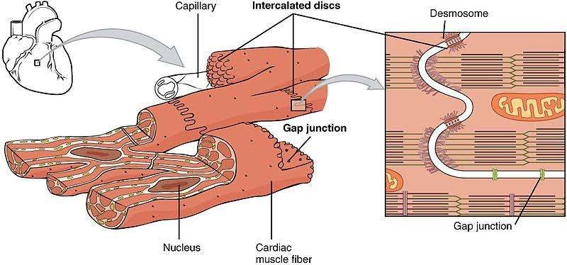
The autonomic nervous system also regulates cardiac muscle, which is located in the heart’s walls. Similar to smooth muscle, the cardiac muscle cell has a single central nucleus, but it is also striated like skeletal muscle. The form of the heart muscle cell is rectangular. Heart muscle contractions are powerful, rhythmic, and involuntary. The heart is the only muscle that never gets tired. It has heart muscle, a particular kind of muscle with a high density of mitochondria. This enables the heart to continue producing energy and pumping blood throughout the body without experiencing weariness.
We shall go into great detail about cardiac and smooth muscle in relation to their respective systems. The skeletal muscle system is the primary topic of this unit. Only located in the heart, cardiac muscle is a striated, specialized, involuntary muscle. To move blood throughout the body, it contracts in a rhythmic manner.
Essential Terms
- Cardiomyocyte: Heart muscle cell
- Intercalated discs: Cell junctions
- Sarcomere: A contraction’s functional unit
- Autorhythmicity: The capacity to contract in the absence of outside stimuli
- Syncytium: A functional unit in which cells cooperate
Muscle Interaction
- Heart muscle functions as a single, cohesive unit.
- Intercalated discs connect cells, enabling quick signal transmission.
- Guarantees that the heart contracts in unison.
Anatomy of Cardiac Muscle
- Branched, short fibers
- One central nucleus, occasionally two. Packed with mitochondria. Intercalated discs (gap junctions + desmosomes) connect them.
Actin, a thin filament
- composed of the protein actin
- includes tropomyosin and troponin.
- aids in controlling contraction
Myosin, or thick filament
- composed of the protein myosin
- creates cross-bridges with actin.
- Generates the force of contraction
Nerve and Blood Supply
Supply of nerves:
- The autonomic nervous system
- The sympathetic nervous system raises heart rate
- The parasympathetic nervous system lowers heart rate.
Blood supply:
- Supplied via the coronary arteries
- High requirement for oxygen
Heart Muscle Structure
Like skeletal muscle, striated branching cell network. The myocardium, or heart wall, is formed of intercalated discs for the connection.
Contraction of Cardiac Muscle
- Self-exciting, or myogenic
- SA node (pacemaker) control
- Makes use of calcium-induced calcium release
- Prevents tiredness due to its extended refractory period.
- The sliding filament mechanism is followed by contraction.
Heart Muscle Functions
- Circulates blood throughout the body.
- Keeps the blood flowing.
- Aids in controlling blood pressure
- Continually works without getting tired
Smooth Muscles
The autonomic nervous system regulates smooth muscle, which is present in the walls of hollow internal organs such as the uterus, bladder, gastrointestinal tract, and blood vessels. Smooth muscle operates involuntarily since it cannot be controlled intentionally. The smooth, non-striated muscle cell has a single central nucleus and is shaped like a spindle. Smooth muscle contracts rhythmically and slowly. The walls of hollow organs like the intestines, blood vessels, uterus, and bladder include smooth muscle, which is an involuntary, non-striated muscle. It controls bodily functions on its own.
EssentialTerms
- Smooth muscle cell, or myocyte: Spindle-shaped cells have a single nucleus and tapered ends.
- Dense bodies behave like attachment points, or Z-lines.
- Peristalsis: a contraction movement that resembles waves. Internal organs contain visceral muscle.
Muscle Interaction
- Coordinated clusters of smooth muscle are seen in both longitudinal and circular strata.
- For instance, the movement of the intestines (peristalsis).
- The lumen narrows when the circular layer compresses.
- Tube shortening due to longitudinal layer contraction.
Anatomy of Smooth Muscle
- Not striated (no bands)
- Spindle-shaped cells
- One central nucleus
- Arranged in layers or sheets
- Has thick bodies rather than sarcomeres.
Actin, a thin filament
- Existing in significant quantities
- Affixed to thick substances
- Aids in contraction
Myosin, or thick filament
- Compared to skeletal muscle, it is present but less structured.
- Interacts with actin to cause contraction.
Blood and Nerve Supply
Supply of nerves:
- The autonomic nervous system
- The release of neurotransmitters from varicosities
Blood supply:
- Provided via capillaries
- Less oxygen-demanding than skeletal muscle
Smooth Muscle Structure
- No striations and no sarcomeres
- Unevenly organized filaments
- Gap junctions between cells
- Forms the visceral organs’ walls
Smooth Muscle Contraction
- Slow and involuntary
- Calcium ions (Ca²⁺) cause
- Replaces troponin with calmodulin
- Able to sustain contraction for an extended period of time (latch state)
- Hormones, nerves, and stretch regulate
Smooth Muscle Functions
- Transports materials (food, pee, blood).
- Regulates blood vessel diameter
- Controls the function of organs
- Aids in uterine contractions during labor.
Function
Your body is moved by muscles. They move in two different ways:
- Voluntary movements are under your control. Your muscles move your body to carry out the action you choose to do. Your nervous system manages these movements. Both running around a track and using your phone to browse this page by flicking your thumb are voluntary behaviors.
- Involuntary movements: Without conscious thought, involuntary movements occur spontaneously. Your body’s muscles in and around your organs operate automatically to maintain optimal function. Examples of involuntary movements include your heartbeat and the muscles in your back and chest that move your ribs when you breathe.
Your muscles interact with almost every system and function in your body through a combination of voluntary and involuntary movements. Different muscle types help with:
- Vision.
- Listening.
- Speaking, swallowing, and breathing.
- Breaking down meals and getting rid of waste (urinating and defecating).
- Standing up straight, sitting still, and moving.
- Circulating blood via your heart and blood vessels.
- Giving birth.
- Additionally, as part of your metabolism, muscles collect and release energy.
FAQs:
What are the 3 main muscle types?
The human body contains three main types of muscle: smooth, cardiac, and skeletal. Every type of muscle has distinct physiology, pathologies, cellular components, and functions. Skeletal muscle is one organ that primarily controls posture and movement.
What is the strongest muscle in the body?
The masseter, the main chewing muscle, is the strongest muscle by weight. When all of the jaw’s muscles cooperate, it can close the teeth with a force of up to 200 pounds (90.7 kilograms) on the molars and 55 pounds (25 kilograms) on the incisors.
What is the weakest human muscle?
The stapedius muscle in the middle ear is thot to be the weakest in the human body. Its length is only about 1 millimeter, and its cross-sectional area is roughly 0.5 mm 2.
Which muscle never gets tired?
The heart is the one muscle that never gets tired. It contains heart muscle, a specialized type of muscle with high mitochondrial density. This enables the heart to continue producing energy and pumping blood throughout the body without experiencing weariness.
How many muscles does the human body have?
Your body has about 600 muscles that are found practically everywhere.
The biceps in your upper arms or the quadriceps in your thighs may be the first muscles that come to mind when you think about muscles, but practically every portion of your body is supported by muscles.
Is a muscle a tissue or an organ?
Tissue makes up muscles. Some of your organs are lined by smooth muscle tissue, but the majority of your organs are composed of different kinds of tissue. Healthcare professionals sometimes refer to the muscular system as a single anatomical group comprising all your muscles. They may also incorporate muscles as components of other bodily systems.
The only organ that is also a muscle is your heart. It is composed of heart muscle, a unique kind of muscle tissue. The only organ in your body that contains cardiac tissue is your heart.
Reference:
- Professional, C. C. M. (2025b, December 29). Muscles. Cleveland Clinic. https://my.clevelandclinic.org/health/body/21887-muscle
- Muscle types | SEER training. (n.d.). https://training.seer.cancer.gov/anatomy/muscular/types.html
- Structure of skeletal muscle | SEER training. (n.d.). https://training.seer.cancer.gov/anatomy/muscular/structure.html
- Introduction to the muscular system | SEER training. (n.d.). https://training.seer.cancer.gov/anatomy/muscular/