Muscular System Explained: Types, Function, Anatomy
What is the Muscular System?
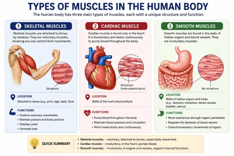
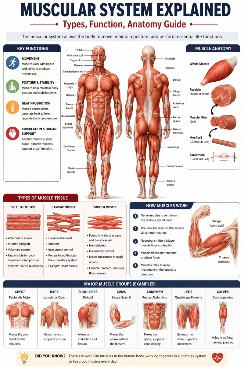
All of the body’s muscles make up the muscular system. Skeletal muscles, which are connected to bones and allow for voluntary bodily motions, make up the majority of muscles in the muscular system.
The human body contains around 650 skeletal muscles, several of which are seen in the figure.In addition to skeletal muscles, the muscular system also consists of smooth muscles, which regulate movement in various interior organs and structures, and cardiac muscle, which forms the heart’s walls.
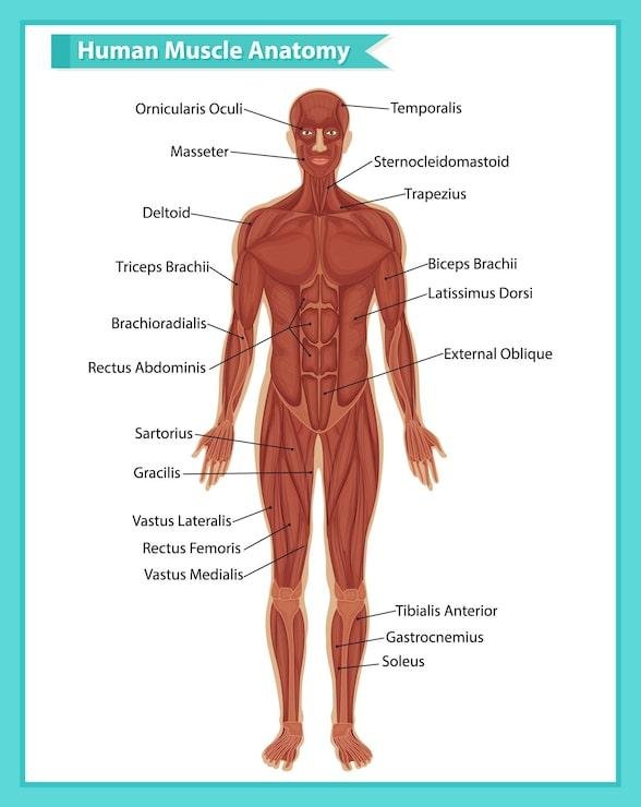
Each muscle is made up of hundreds or tens of thousands of tiny muscle fibers that form a type of elastic tissue. Each muscle fiber is made up of several microscopic strands called fibrils, and each muscle fiber’s contraction is controlled by nerve cell impulses.
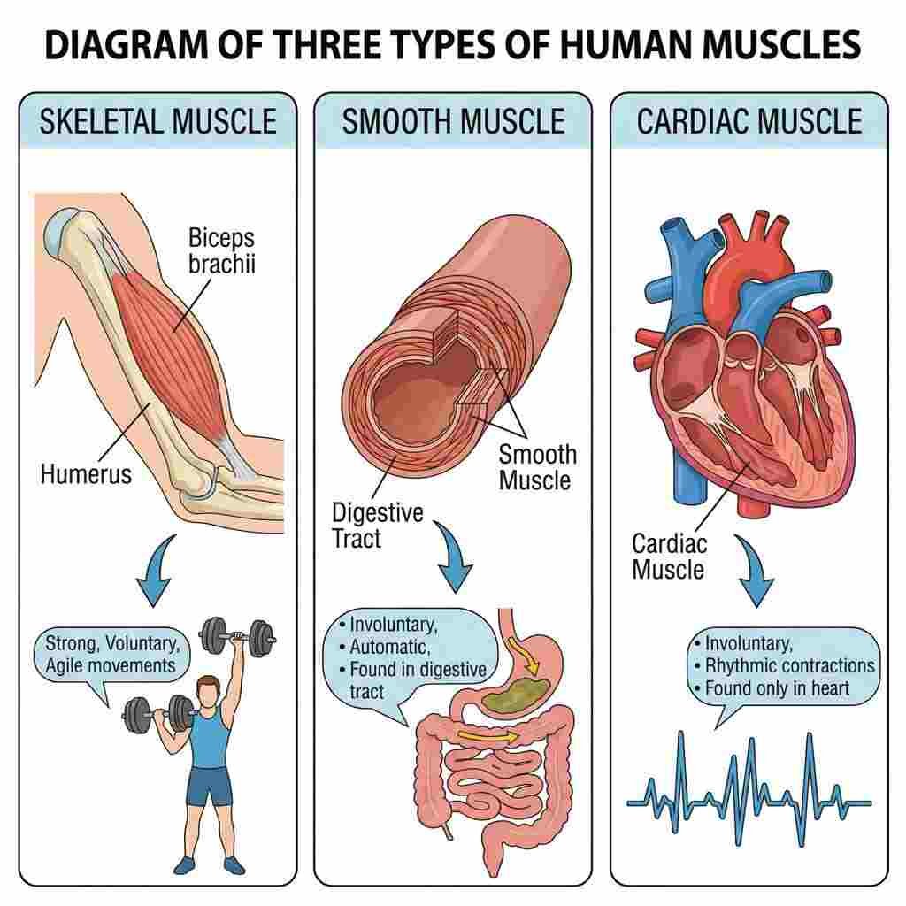
Types of muscles
Three different kinds of muscle. The strength, balance, posture, mobility, and the body’s ability to stay warm are all provided by muscles.
- Skeletal muscle
- Cardiac or heart muscle
- Smooth (non-striated) muscle
Skeletal muscle
Skeletal muscle is a kind of striated muscle made up of muscle fibers, which are made up of myofibrils. Sarcomeres, the fundamental units of striated muscle tissue, make up myofibrils. Skeletal muscles shorten each sarcomere in a coordinated contraction when stimulated by an action potential.
The sliding filament model of muscle contraction is the most well-proposed model for comprehending contraction. Actin and myosin fibers overlap in a contractile action toward one another within the sarcomere. The club-shaped myosin heads of myosin filaments extend in the direction of the actin filaments and give the actin filaments places to adhere to binding sites. The myosin heads rotate in the direction of the sarcomere’s center, separate, and then reconnect to the actin filament’s closest active site in a coordinated manner. We refer to this as a ratchet-type drive mechanism.
Adenosine triphosphate (ATP), the cell’s energy source, is heavily used in this process. Actin filaments and myosin heads form cross-bridges that are bound by ATP. The myosin head rotates due to the release of energy. Adenosine diphosphate (ADP) is created when ATP is consumed, and because muscles cannot store much ATP, they must constantly replenish the released ADP with ATP. Additionally, muscle tissue has a reserve of creatine phosphate, a fast-acting recharge molecule that may help quickly regenerate ADP into ATP as needed.
Every sarcomere cycle requires calcium ions. When a muscle is contracted, calcium is released from the sarcoplasmic reticulum into the sarcomere. The actin-binding sites are revealed by this calcium. The calcium ions are pushed out of the sarcomere and back into storage in the sarcoplasmic reticulum when the muscle is no longer required to contract.
The skeleton’s bones are connected to skeletal muscles. The body moves when these muscles flex. They enable us to do many tasks with our limbs, such as walking and cartwheeling. Additionally, skeletal muscles support balance and posture.
Cardiac or heart muscle
The cardiac muscle (myocardium), which is made up of individual cardiac muscle cells connected by intercalated discs and surrounded by collagen fibers and other materials that form the extracellular matrix, is the primary tissue of the heart wall. It is an involuntary, striated muscle that forms a thick middle layer between the inner layer (the endocardium) and the outer layer (the pericardium), with blood supplied via the coronary circulation.
Only the heart’s walls contain cardiac muscle tissue. The heartbeat is produced by the contraction of cardiac muscle. The circulatory system’s blood flow is maintained by the heart’s pumping function.
Smooth (non-striated) muscle
It is regulated by the autonomic nervous system and is present in invertebrates as well. Because it lacks sarcomeres and thus striations (bands or stripes), it is referred to as non-striated.Single-unit and multi-unit smooth muscle are its two subgroups. The whole bundle or sheet of smooth muscle cells in a single-unit muscle contracts as a syncytium.
The walls of hollow organs, such as the stomach, intestines, bladder, and uterus, include smooth muscle. Vascular smooth muscle is found in the walls of blood arteries and lymph vessels (apart from blood and lymph capillaries). The reproductive, urinary, and respiratory tracts include smooth muscle. Smooth muscles in the eyes include the iris dilator, iris sphincter, and ciliary muscles. The pupils can be dilated or constricted by contracting the iris’s sphincter and dilator muscles. In order to concentrate on objects in accommodation, the ciliary muscles alter the curvature of the lens. In reaction to anxiety and cold temperatures, smooth muscle cells in the skin, such those of the arrector pili, cause hair to stand upright.
Vasoconstriction, which results from the contraction of smooth muscles in blood vessel walls, may aid in the preservation of body heat. Vasodilation brought on by the relaxation of these muscles may aidin the body’s heat loss. Smooth muscles of the digestive system’s organs contract sequentially to create a wave of muscular contractions known as peristalsis, which forces food through the gastrointestinal tract. You can get a decent picture of how muscles transport food through the digestive system by picturing yourself squirting toothpaste through a tube by applying pressure sequentially from the bottom to the top. Urine is also moved along the urinary system by smooth muscle movement.
Functions of muscle tissue
- Movement: Walking, eating, running, lifting, using our hands to manipulate items, and picking our noses are all made possible by the rigidity of our skeleton, which allows skeletal muscles to pull on it.
- Posture maintenance: Our muscles produce a continuous contractile force that enables us to maintain an upright or sitting position, or posture, without much conscious control.
- Respiration: Air is naturally forced into and out of our bodies by our muscular system. Heat generation: Heat is produced as muscle tissue contracts, and this heat is necessary to maintain temperature homeostasis. For example, we shudder to produce more heat when our core body temperature drops.
- Communication: Muscle tissue enables us to write, speak, gesture, and express our emotions through facial expressions like frowning or smiling.
- Organ and blood vessel constriction: Smooth muscle contraction forces secretions out of glands, nutrients travel through our digestive tract, and urine exits the body. Blood pressure and blood distribution throughout the body are controlled by the constriction or relaxation of blood vessels.
- Pumping blood: Because our heart constantly takes in blood and distributes it to all of the body’s tissues and organs, blood flows via the blood vessels.
This is not an exhaustive list. The fact that muscles are essential for preserving the integrity of bodily cavities and for protecting delicate inside organs by enclosing them are only two of the numerous instances. For instance, belly contents herniate (protrude) up into the thoracic cavity in fetuses with inadequate diaphragms, impeding normal lung growth and development. Although this is not a comprehensive list, understanding some of these fundamental muscle activities will be helpful to you as we move on.
Properties of muscle tissue
Excitability, Contractility, extensibility, and elasticity are characteristics that all muscle cells have in common. All muscle tissue is made up of “muscle fibers,” which are muscle cells with certain properties like the following:
- Excitability: the ability to react to stimuli by starting an electrical signal (action potential) that travels across the plasma membrane and sets off internal processes that cause muscles to contract.
- Contractility: cell shortening due to contraction.
- Extensibility: the ability to lengthen in reaction to opposing muscle cells contracting.
- Elasticity: the capacity to revert to its initial length upon the removal of tension.
Muscle Hypertrophy and Atrophy
Hypertrophy is the expansion of muscles. Increased usage is typically the cause of this; however, hormonal or other factors may also be involved. For instance, a notable increase in muscular growth is brought on by the rise in testosterone throughout puberty. Almost everyone may improve the size of their skeletal muscles with weight-bearing or resistance training exercises. Exercises that raise heart rate, like jogging, may also increase cardiac muscle growth and strength. In turn, muscle size is the primary factor that determines muscular strength, which may be quantified by the force that a muscle can produce.
Atrophy, or the shrinking of muscles, can also happen as a result of hunger or inactivity. People who are immobile for an extended period of time—for instance, due to surgery or a broken bone—lose muscle mass somewhat fast. People in concentration or famine camps may be so undernourished that they essentially become “skin and bones,” losing much of their muscular mass. Because they are weightless in orbit, astronauts aboard the International Space Station may also lose a considerable amount of muscular mass.
Muscle atrophy is frequently linked to many illnesses, such as AIDS and cancer. Age-related muscular atrophy also occurs. Sarcopenia is the term for the progressive loss of skeletal muscle mass that occurs as people age. Although the precise origin of sarcopenia is unknown, a reduction in sensitivity to growth factors—which are essential for maintaining muscle mass—is one potential explanation. Muscular atrophy results in a commensurate decrease in muscular strength since muscle size dictates strength.
The quantity of muscle fibers remains constant in both hypertrophy and atrophy. What causes the muscle fibers to alter in size? Muscle hypertrophy causes the individual fibers to widen. The fibers get narrower as muscle atrophy occurs.
Relationships with Other Systems of the Body
Muscles are unable to contract by themselves. For skeletal muscles to contract, motor neurons must stimulate them. A neuromuscular junction is the location where a motor neuron connects to a muscle. Suppose you choose to raise your hand during class. Your arm and shoulder get electrical signals from your brain via motor neurons. Your arm rises as a result of the motor neurons stimulating the muscle fibers in your shoulder and arm to contract.
Electrical impulses also regulate the involuntary contractions of smooth and cardiac muscles, although in the case of these muscles, the impulses originate from either specific cells in the heart (cardiac muscle) or the autonomic nervous system (smooth muscle). Involuntary contractions of the heart and smooth muscles are also influenced by hormones and a few other variables. For instance, the fight-or-flight hormone adrenaline speeds up the heartbeat by increasing the pace at which cardiac muscle contracts.
The body cannot be moved by muscles alone. To take action, they require the skeletal system. The term “musculoskeletal system” is frequently used to refer to the two systems together. Tough connective structures called tendons hold skeletal muscles to the skeleton. Numerous skeletal muscles are joined to the ends of bones at joints. The muscles link the bones and span the joint. The bones move as a result of the muscles pulling on them as they contract. Body movement is made possible via a series of levers provided by the skeletal system. The force used to move the levers comes from the muscular system.
Clinical significance
Muscular dystrophy
A collection of conditions known as muscular dystrophy is linked to increasing muscle weakening and muscle mass loss. Genetic mutations are the cause of several illnesses. The illness affects 19.8 to 25.1 people per 100,000 person-years worldwide.
Muscular dystrophy comes in more than thirty varieties. Muscular dystrophy can impact a person’s heart and lungs, as well as their capacity to walk, move, and carry out everyday tasks, depending on the kind. The most prevalent kinds consist of:
- Becker and Duchenne muscular dystrophy
- Myotonic dystrophy
- Muscular dystrophy of the limbs and girdles
- Dystrophy of the facioscapulohumeral
- Dystrophy congenital
- Distal
- Muscular dystrophy of the oculopharynx
- Emery-Dreifuss muscular dystrophy
Myopathy
Myopathy is a medical term for a muscular illness when the muscle fibers malfunction. Myopathy is Greek for “muscle disease” (myo-muscle + patheia-pathy: pain). This interpretation suggests that the main flaw is in the muscle rather than the nerves (“neuropathies” or “neurogenic” illnesses) or another organ (such as the brain).
Myalgia (muscle discomfort), muscle weakness (reduced muscle force), or premature muscle exhaustion (initially normal but diminishing muscle force) are the usual outcomes of this muscular abnormality. Myopathy may also be linked to contracture, spasm, stiffness, and cramping of the muscles. Long-term (chronic) myopathy can cause a muscle to develop an aberrant size, such as muscular atrophy (abnormally tiny) or a pseudoathletic look (abnormally huge).
Capture myopathy causes morbidity and mortality in both wild and confined animals, including kangaroos and deer. Stress and physical strain during capture and restraint are typically the causes.
There are two types of muscle diseases: neuromuscular and musculoskeletal. Various myopathies might be idiopathic (cause unknown), infectious, hereditary, or non-communicable. As is common with mitochondrial myopathies, the illness may be isolated to impact only muscle (pure myopathy), or it may be a component of a systemic illness.
Symptoms and indicators: Muscle weakness, cramping, stiffness, and tetany are typical symptoms.
Diagnosis: Since no single test can diagnose every kind of myopathy, the examination of a suspected myopathy includes laboratory, electrophysiological, imaging, histological, and genetic testing in addition to clinical assessment.
Muscle biopsy
A muscle biopsy is a medical process where an organism’s muscle tissue is taken out and studied under a microscope. Issues with the neurological system, connective tissue, vascular system, or musculoskeletal system may be found with a muscle biopsy.
Symptoms and indicators: A muscle biopsy can help differentiate between neuropathies (where the pathology is at the nerves innervating those muscles) and myopathies (where the disease is in the muscle tissue itself) in persons with weakness and poor muscle tone. By examining muscle samples under a microscope for distinct features when subjected to a range of chemical reactions and stains, it is also possible to differentiate between different kinds of myopathies.
Sometimes, nevertheless, a muscle biopsy is not enough to differentiate between different myopathies. For instance, “centronuclear myopathy” would be indicated by a muscle biopsy that showed the nucleus pathologically positioned in the center of the muscle cell. However, research has shown that a variety of myopathies can cause these centronuclear biopsy appearances, so specific genetic testing becomes more crucial. Furthermore, the only reliable method to identify the different kinds of muscle fibers is by muscle biopsy. For example, by having a muscle biopsy, one may clearly see what kind of muscles predominate in their body.
Diagnosis: Typically, a biopsy needle is inserted into a muscle, leaving behind a little bit of tissue. An “open biopsy” is an alternative method that involves making a tiny surgical incision to remove the muscle tissue.
FAQs
What are the main types of muscles?
Skeletal, smooth, and cardiac muscles are the three primary categories of muscle.
What is the structure of the muscle?
A layer of connective tissue known as the perimysium envelops each bundle of muscle fibers, known as a fasciculus. The endomysium, a type of connective tissue, envelops each individual muscle cell, also known as a muscle fiber, within the fasciculus.
What are four disorders of the muscular system?
Muscular dystrophy (diseases that weaken muscles), tendinosis (degenerative tendon disease), fibromyalgia (chronic pain), mitochondrial myopathy (mitochondrial ATP disorder), myasthenia gravis (immune system issue), and tetanus (paralyzing bacterial infection) are examples of diseases and disorders of the muscular system.
References
- Lutz, G. J., & Rome, L. C. (1994). Built for Jumping: the Design of the Frog Muscular System. Science, 263(5145), 370–372. https://doi.org/10.1126/science.8278808
- Spudich, J. A., & Watt, S. (1971). The regulation of rabbit skeletal muscle contraction. Journal of Biological Chemistry, 246(15), 4866–4871. https://doi.org/10.1016/s0021-9258(18)62016-2
- Noma, A. (1983). ATP-regulated K+ channels in cardiac muscle. Nature, 305(5930), 147–148. https://doi.org/10.1038/305147a0
- Furchgott, R. F., & Zawadzki, J. V. (1980). The obligatory role of endothelial cells in the relaxation of arterial smooth muscle by acetylcholine. Nature, 288(5789), 373–376. https://doi.org/10.1038/288373a0
- Jackson, S. P., & Bartek, J. (2009). The DNA-damage response in human biology and disease. Nature, 461(7267), 1071–1078. https://doi.org/10.1038/nature08467
- Duan, D., Goemans, N., Takeda, S., Mercuri, E., & Aartsma-Rus, A. (2021). Duchenne muscular dystrophy. Nature Reviews Disease Primers, 7(1), 13. https://doi.org/10.1038/s41572-021-00248-3