Optic Nerve
Introduction
The optic nerve is a vital nerve that connects the eye to the brain and carries visual information from the retina to the brain for processing.
The back of the eye is where the optic nerve is located. The second cranial nerve, often known as cranial nerve II, is one of multiple pairs of cranial nerves. It is a nerve cell bundle that sends electrical impulses from the eye to the brain to provide sensory information for vision. The optic nerve has been extensively investigated since it is a direct extension of the brain.
It plays a key role in vision, helping us recognize shapes, colors, movement, and details in our surroundings.
Anatomy
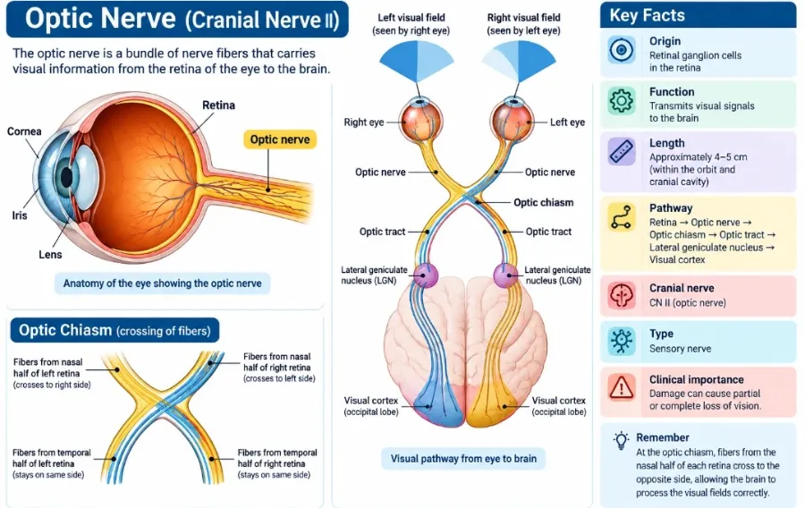
The retinal ganglion cells’ axons (nerve fibres) make up the majority of the optic nerve. The optic disc, also known as the nerve head, is where the axons of the retinal ganglion cells leave the eye.
The nerve head is seen as a white circular structure at the back of the eye. There are no photoreceptors in this structure. As a result, humans possess a natural blind zone.
Nerve cells exit the nerve head via a structure known as the lamina cribrosa, which permits nerve fibres to pass through many pores and enter the extraocular (outside of the eyeball) region. As the fibres travel through, they are wrapped with a sort of insulation known as myelin. Oligodendrocytes, a kind of glial cell, insulate nerve fibres.
Location
The retinal ganglion cells’ axons (nerve fibres) make up the majority of the optic nerve. The optic disc, also known as the nerve head, is where the axons of the retinal ganglion cells leave the eye.
The nerve head is seen as a white circular structure at the back of the eye. There are no photoreceptors in this structure. As a result, humans possess a natural blind zone.
Nerve cells exit the nerve head via a structure known as the lamina cribrosa, which permits nerve fibres to pass through many pores and enter the extraocular (outside of the eyeball) region. As the fibres travel through, they are wrapped with a sort of insulation known as myelin. Oligodendrocytes, a kind of glial cell, insulate nerve fibres.
Pathway
The optic tract is an intracranial extension of the optic nerve. The optic tract, like CN II, is divided into pairs. Each is made up of temporal fibres from the ipsilateral retina and nasal fibres from the contralateral retina. The majority of these fibres will go to the thalamic lateral geniculate body and connect with the dorsal lateral geniculate nucleus.
However, a subset of these fibres will skip the lateral geniculate body and end in the pretectal nucleus (participating in the pupillary light reflex) and superior colliculus (regulating saccadic eye movements). However, fibres escape the lateral geniculate body and create the optic radiation (geniculocalcarine tract). The fibers leave the lateral geniculate body through the retrolenticular area of the internal capsule, giving rise to the six layers that make up this tract. The fibres eventually end in Brodmann area 17 (the visual cortex) of the calcarine sulcus.
Pupillary light reflex.
The pupillary light reflex refers to the reflex that governs the diameter of the pupil. The optic nerve relays information to the pretectal nucleus in the superior colliculus, which regulates the activity of the Edinger-Westphal nucleus (CN III’s parasympathetic nucleus). The fibres of the optic tract that connect to the pretectal nucleus do not interact with the lateral geniculate body.
The presynaptic parasympathetic fibres of each Edinger-Westphal nucleus then go with the oculomotor nerve fibres to the ciliary ganglion, where they synapse. The postsynaptic parasympathetic fibres then move via the short ciliary nerve to innervate the iris’ constrictor pupillae. As a result, the pupillary light reflex route consists of one afferent limb from the ipsilateral optic tract and two efferent limbs that give bilateral innervation to the Edinger-Westphal nuclei. As a result, it can induce both direct (ipsilateral pupillary constriction in response to a light stimulus) and consensual (contralateral pupillary constriction in response to a light stimulus) pupillary responses.
Optical radiation (geniculocalcarine tract)
This dorsal lateral geniculate nucleus is split into layers 1–6, with a somatotopic layout in which fibres from the contralateral eye synapse on layers 1, 4, and 6. Conversely, fibres from the ipsilateral eye will synapse on layers 2, 3, and 5. Axons from the upper quadrants (except the macula) also project to the anteromedial nucleus. Similarly, those originating in the inferior quadrants (excluding the macula) project to the anterolateral region of the nucleus. The upper and lower quadrants of the macula will project to the posteromedial and posterolateral regions of the lateral geniculate nucleus, respectively.
The fibres leaving the lateral geniculate body are collectively referred to as the geniculocalcarine tract. They carry integrated visual impulses from the thalami to the visual cortex and are separated into upper and lower loops. The upper loop fibres go far anteriorly across the temporal horn of the lateral ventricle, passing through the temporal lobe, before bending posteriorly into the calcarine sulcus.
The fibres of the lower loop travel directly through the parietal lobe and into the retrolentiform region of the internal capsule. Fibres from the peripheral upper and lower quadrants eventually project to the anterior two-thirds of the primary visual cortex, whilst those from the upper and lower central quadrants (i.e. the macula) project to the posterior third of the main visual cortex.
Frontal eye fields.
In addition to the various thalamocortical projections related to vision, postsynaptic fibres from the main visual field proceed to the cerebrum to synapse and integrate with the frontal eye fields (Brodmann 6, 8, and 9). This part of the brain contains several connections to the thalamus, as well as the parietal and temporal lobes, where it processes afferent information relating to sight and hearing.
The frontal eye fields, via their efferent fibres to the ipsilateral Edinger-Westphal and primary oculomotor nuclei, can govern fast eye movements between fixation locations.
Blood supply
The blood flow to the optic nerve varies depending on the section being discussed. In essence, the internal carotid artery’s ophthalmic branch supplies the optic nerve indirectly. The internal carotid artery exits superiorly from the cavernous sinus, whereas the ophthalmic artery diverges and travels along the ventral side of the optic nerve within the optic canal. As the artery exits at the top of the orbit, it creates three posterior ciliary arteries, as well as the central retinal artery. After piercing the structure at 10 – 12 mm from the globe, the veins proceed anteriorly into the optic nerve’s substance.
The surface nerve fibre layer of the optic nerve head is perfused by branches of the central retinal artery. The prelaminar zone receives blood flow via peripapillary and choroid vessels that branch from the posterior ciliary arteries. The blood supply to the lamina cribrosa comes from the small posterior ciliary arteries and, in certain circumstances, the Zinn-Haller circle. The latter vascular structure is variable; however, it can be found in the subscleral area surrounding the optic nerve at the neuro-ocular junction. Tributaries to the Zinn-Haller network are formed by the choroidal vessels, pial arterial network, perineural arteries, and around 4 – 8 posterior ciliary arteries. The Zinn-Haller vascular network also branches into the laminar region of the optic disc.
The posterior ciliary arteries supply the orbital section of CN II through the pial vessels, which connect to the nerve via fibrous septa. The internalised portion of the central retinal artery may also give arterial feed to the nerve here. The intraorbital CN II at the orbital apex may get collateral blood flow from the external carotid artery’s middle meningeal branch as well.
The intracanalicular portion of CN II is fed by the collateral ophthalmic artery (anteriorly) and pial derivatives of the superior hypophyseal and internal carotid arteries (posteriorly). The superior hypophyseal, internal carotid, and A1 part of the anterior cerebral arteries supply CN II within the cranial vault. Although the optic nerve is classified as a diencephalic extension, the supplying arteries lack a blood-brain barrier.
Venous tributaries from all areas of the optic nerve eventually flow into the central retinal vein. It subsequently empties into the superior ophthalmic vein or straight into the cavernous sinus.
Function
The optic nerve generates all types of visual information.
The optic nerve allows humans to see brightness, colour, and contrast.
The optic nerve also controls the light reflex and the accommodation reflex.1These are two key neurological responses. When light enters one of the eyes, the light reflex causes both pupils to constrict. The accommodation reflex helps the eye to compensate for close vision by causing the lens to enlarge.
Related condition
Several illnesses can impact the optic nerve, chiasma, and radiations, including:
Glaucoma
Glaucoma is a category of disorders that can cause damage to the optic nerve. Optic nerve fibres are a component of the retina that allows humans to see. This nerve fibre layer can be destroyed if the eye’s pressure (intraocular pressure) becomes too high.
High pressure eventually causes nerve fibres to die, resulting in impaired eyesight. Glaucoma is likely to result in blindness and vision loss if treatment is not received.
Optic Neuritis
An inflammation of the optic nerves is known as optic neuritis. This usually only affects one eye at a time and damages the nerve just before the optic chiasm. Because of the location of the inflammation, one would expect issues with vision in only one eye.
Multiple sclerosis,4 a viral infection, chemical exposure, or severe sinus disease are all potential causes of optic neuritis.
Common signs of optic neuritis:
- Eye discomfort.
- Vision impairment.
- Symptoms may include increased light sensitivity, visual loss, and blind patches.
Pituitary Adenoma
The pituitary gland is placed below the optic chiasm. If the pituitary gland expands or develops a mass or growth, it can impinge on the optic chiasm, creating abnormalities in both visual fields since nerve fibres cross at the chiasm.
Vascular infarctions and aneurysms
Vascular illnesses (diseases that damage blood vessels) can interfere with the optic radiation pathway because the optic radiation nerve fibres run via the brain’s parietal, temporal, and occipital lobes; deficiencies or blind spots in the visual field may emerge. Doctors can use the location of the visual field defect to determine where the issue is in the brain.
Treatment
The treatment of optic nerve, chiasma, and optic radiation damage is determined by the aetiology. However, therapies for optic nerve injury may not restore lost vision. In most situations, precautions are taken to prevent additional harm and worsening of symptoms. For example:
Glaucoma is caused by increasing pressure inside the eye; hence, drugs for glaucoma are designed to reduce the pressure to the point where the disease process stops. Although glaucoma can be treated with surgery, lasers, and oral drugs, the majority of cases are treated with topical treatment in the form of eye drops.
Oral and intravenous steroids are used to treat diseases like optic neuritis, which cause inflammation. Additionally, if the source of the optic neuritis is identified, the underlying problem will be addressed.
Diseases of the optic chiasm are frequently treated with neurosurgery and medicine or hormones. Depending on the severity of optic chiasm illness, such as a pituitary adenoma, mere observation may be sufficient.
Vascular accidents, or strokes, are more difficult to treat unless the issue is detected immediately. Occasionally, blood thinners are given. Surgery may be required if the condition is caused by aneurysms.
FAQs
Can optic nerve damage be treated?
There are no effective treatments to regenerate nerve cells or to restore connections between the eye and brain once the optic nerve is lost.
What vitamin is good for the optic nerve?
Iron, thiamin, and B vitamins are also vital. A thiamin shortage can occur rapidly and affect the optic nerve and eye movement. Non-starchy plant foods, such as fruits and leafy vegetables, are ideal sources of vitamin B
What is the latest optic nerve treatment?
The researchers were able to induce nerve cells in the severed optic nerves of mice to regenerate from the injured area all the way to the optic chiasm in the brain by using an injectable peptide, which is a tiny fragment of a bigger protein.
References:
- Bedinghaus, T., OD. (2023, May 28). The anatomy of the optic nerve. Verywell Health. https://www.verywellhealth.com/optic-nerve-anatomy-4686150
- Optic nerve. (2023, August 16). Kenhub. https://www.kenhub.com/en/library/anatomy/the-optic-nerve