Plantaris Muscle: Anatomy, Function, Injury, and Rehabilitation
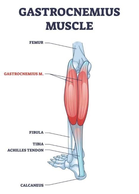
The plantaris is a thin, elongated muscle located in the posterior lower leg, extending from the area behind the knee into the calf region. Along with the gastrocnemius and soleus, it forms the superficial group of the posterior compartment of the leg.
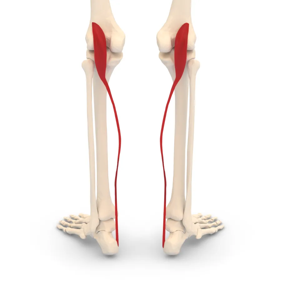
It has a thin muscle belly and a long, thin tendon. The belly is only about 7 to 10 cm long, and its size varies from person to person. The tendon is the longest in the human body, usually 30 to 45 cm (12 to 18 inches), but it is much thinner than the Achilles tendon. In about 10% of people, the plantaris is missing altogether, and sometimes the muscle has two bellies separated by a tendon.
The name comes from the fact that in many other mammals the muscle inserts into the plantar aponeurosis. In humans it doesn’t reach that far. Because it crosses both the knee and the ankle, it can assist knee flexion and plantar flexion, but only weakly.
Origin
The plantaris begins at the lower end of the lateral supracondylar line of the femur, just above the lateral head of the gastrocnemius. The attachment often extends into the oblique popliteal ligament. The tendon then runs down and inward, between the gastrocnemius and soleus. It is so thin that first-year students often mistake it for a nerve during dissection, which is why it’s called the “freshman’s nerve.”
Insertion
The plantaris tendon travels along the inner side of the Achilles tendon, which is formed by the gastrocnemius and soleus muscles, before inserting into the posterior surface of the calcaneus (heel bone). Sometimes it blends with the calcaneal tendon, the flexor retinaculum, or the deep fascia of the ankle.
Relations
The plantaris is one of the most superficial muscles of the posterior compartment, and only the gastrocnemius covers it. Along with the lateral head of the gastrocnemius, this muscle contributes to the lower outer boundary of the popliteal fossa. It is positioned superficial to the soleus, with its tendon descending through the space between the gastrocnemius and soleus muscles.
Nerve supply
The plantaris is supplied by the tibial nerve, one of the two main branches of the sciatic nerve. Its fibres come from the S1 and S2 spinal nerves.
Blood supply
The plantaris has two sources of blood supply. Superficially, it receives blood from the lateral sural branch of the popliteal artery. Its deep surface is supplied by the superior lateral genicular artery, which also comes from the popliteal artery. The distal section of the tendon is supplied by small calcaneal branches arising from the posterior tibial artery.
Anatomical variation
The muscle may arise from the oblique popliteal ligament. It frequently blends with the fibres of the lateral head of the gastrocnemius muscle.
Function
There is some debate about what the plantaris actually does. Because of its attachments, it can flex the knee and plantar flex the foot at the ankle. In reality, it only gives slight support to the gastrocnemius and soleus in these two actions.
The plantaris has many muscle spindles, which are proprioceptive receptors, so it has been suggested that it works as a proprioceptive organ for the larger ankle flexors. On the other hand, some studies suggest it may be almost vestigial in humans, since neither flexor strength nor proprioception is affected when it is absent or removed.
Clinical importance
The plantaris is traditionally blamed for a common injury called “tennis leg.” Recent ultrasound studies show that tennis leg is more often caused by a tear at the musculotendinous junction of the medial head of the gastrocnemius, although a plantaris rupture can also cause calf pain. In one clinical study of 141 patients diagnosed with tennis leg, 94 (66.7%) had a partial gastrocnemius rupture, and only 2 (1.4%) had a plantaris tendon rupture.
The injury can happen in sports that need quick push-off from one leg, such as tennis, basketball, and football, during running or jumping. Plantaris ruptures usually occur along with injury to other muscles in the posterior compartment, and isolated strains are rare. A rupture can cause swelling, pain at the back of the lower leg, lingering soreness, and sometimes a pop heard at the time of injury. Moving the ankle, especially stretching it, can also be painful.
Assessment
The muscle belly can be felt in the popliteal fossa, medial to the lateral head of the gastrocnemius and just above it, and along the medial side of the common tendon of the triceps surae. Ask the patient to lie face down and flex the knee until it reaches approximately a right angle (90 degrees). The practitioner rests a forearm against the sole of the foot to resist plantar flexion and knee flexion at the same time, while the other hand palpates the area with the heel supported.
Management
Acute stage
Include the RICE principles in the immediate management:
- Rest
- Ice
- Compression
- Elevation
Research suggests that immobilisation should last no more than one to three days, depending on how severe the injury is, with the foot in a neutral or slightly plantarflexed position.
Sub-acute stage
After immobilisation, progressive passive, active and resisted movements can start within the pain threshold. Manual therapy such as myofascial release, soft tissue mobilisation and active release can also begin at this stage. Manual therapy is important for proper collagen fibre growth and realignment.
Progressive strengthening is also important now. It should stay within the person’s pain threshold and follow the principles of isometric, isotonic and isokinetic training.
Sub-acute to chronic stage
Progressive strengthening and range-of-motion exercises continue. Proprioceptive, balance and sport-specific training can also begin in this phase.
Health tips
Heel pain is the main symptom of plantar fasciitis. It is more common in runners and in people who spend long hours on their feet, but it can affect people of any age and ability.
Research shows that strengthening and stretching exercises, including stretching the Achilles tendon, can reduce pain and improve walking in people with plantar fasciitis.
If you have been diagnosed with plantar fasciitis or have heel pain, a physical therapist can help you plan your treatment. You can also try these six exercises at home.
Exercises
1. Plantar fascia massage
Note: This should not cause pain. Use just enough pressure to feel a mild stretch.
- Sit on a chair or stand with one foot on a small ball or a frozen water bottle. A frozen bottle is helpful because the cold helps reduce inflammation.
- Gently roll the ball or bottle back and forth under your foot, starting just below the ball of your foot and stopping just before the heel.
- Roll back and forth 10 times on each foot. Do 2 sets per foot.
- Do this once a day.
2. Heel raises
Note: Move slowly and carefully. Hold a railing or other support to keep your balance if needed.
- Position yourself on the lowest stair with your heels extending beyond the edge and the front portions of your feet supported on the step.
- Slowly lower your heels just below the edge of the step. You may feel a stretch in your calf muscle.
- Slowly rise back up onto the balls of your feet.
- Do 10 repetitions, rest, then complete the second set.
- Do this once a day.
3. Resisted ankle inversion while sitting on the floor
Note: You need an elastic exercise band for this exercise.
- Sit on the floor with your torso upright, and both legs extended comfortably in front of you. Keep your hips still during the exercise.
- Loop the band around the forefoot of the working foot and hold the other end in your hand. Cross the other leg over so the band stays in place.
- Slowly turn the working foot inward, away from the other foot, then slowly bring it back to the starting position.
- Do 10 repetitions per foot, 2 sets.
- Do this once a day.
4. Toe towel scrunches
Note: Only your toes should work. Keep your whole foot on the floor.
- Sit up straight on a chair, or stand, with one foot on a towel and your toes spread.
- Curl your toes to scrunch the towel and pull it toward you. Do 10 to 15 scrunches per foot, 2 sets.
- As you get stronger and it becomes easier, place a small weight (2 to 4 pounds) on the far end of the towel.
- Do this one to three times a day.
Stretching
1. Seated plantar fascia stretch
Note: Go slowly and stay relaxed.
- Sit in a chair and cross one leg over the opposite knee.
- Hold your ankle with one hand and your toes with the other. Slowly pull your toes back until you feel a stretch under your foot.
- Hold for 20 seconds and repeat 3 times on each foot.
- Do this once a day.
2. Wall calf stretch
Note: Stand tall, facing a wall at arm’s length, with your hands flat on it.
- Step one leg straight back, keeping both feet flat on the floor. Bend your front knee until you feel a stretch in the calf of the back leg.
- Hold for 20 seconds and repeat 3 times on each leg.
- Do this once a day.
FAQs
What is the nerve root of the plantaris?
The tibial nerve supplies neural innervation to the plantaris muscle (S1, S2).
What supplies the plantaris muscle?
The plantaris has two sources of blood. The lateral sural artery, a branch of the popliteal artery, provides blood to it superficially. The popliteal artery also gives rise to the superior lateral genicular artery, which supplies its deep surface.
What is the plantaris muscle used for?
The ankle and knee joints are weakly plantar flexed by the plantaris. The central nervous system may also receive proprioceptive signals about the foot’s location from the plantaris muscle. This idea is supported by the exceptionally high density of proprioceptive receptor end organs.
What does plantaris pain feel like?
The most common symptoms are calf and behind-the-knee pain. Pain typically starts as soon as an injury occurs. When the damage first happens, some people may also experience a sudden, excruciating pain in the affected location. Ankle mobility may also be challenging in some situations.
References:
- Wikipedia contributors. (2024, May 4). Plantaris muscle. Wikipedia. https://en.wikipedia.org/wiki/Plantaris_muscle
- Health Tips | Six exercises for plantar fasciitis and heel pain. (2024, March 26). Choose PT. https://www.choosept.com/health-tips/six-exercises-plantar-fasciitis-heel-pain
- Plantaris muscle. (2023, October 30). Kenhub. https://www.kenhub.com/en/library/anatomy/plantaris-muscle
- vastralphysiotherapyclinic. (2023). Plantaris Muscle Origin, Insertion, Function, Exercise. Mobility Physiotherapy Clinic. https://mobilephysiotherapyclinic.net/plantaris-muscle/