Descending Aorta
Introduction
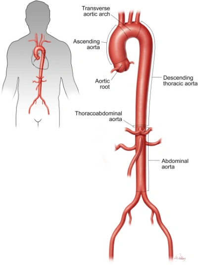
The aorta is the body’s largest artery, carrying oxygenated blood from the heart to supply organs and tissues throughout the body. It originates in the left ventricle and is anatomically divided into five regions: the aortic root, ascending aorta, aortic arch, descending thoracic aorta, and abdominal aorta. As it reaches the lower abdomen, the abdominal aorta separates into the right and left common iliac arteries, which supply blood to the pelvis and lower limbs.
The thoracic aorta is the section of the aorta that extends through the chest cavity. Starting from the aortic arch, it descends close to the left side of the vertebral column before passing through an opening in the diaphragm called the aortic hiatus. Beyond this point, it becomes the abdominal aorta. The vessel’s diameter is not uniform throughout its course and can differ depending on the location and the individual.
The thoracic aorta gives rise to several branches, including the bronchial, esophageal, mediastinal, pericardial, superior phrenic, and posterior intercostal arteries. Typically, it gives off nine pairs of posterior intercostal arteries that supply the chest wall and intercostal muscles. Since the aorta lies to the left of the vertebral column, its right-sided branches are generally longer than its left-sided branches.
Through these branches, the thoracic aorta supplies oxygenated blood to various structures in the chest, including the bronchi, esophagus, mediastinum, chest wall, ribs, intercostal muscles, and parts of the diaphragm.
What Is the Descending Aorta?
The descending aorta is the part of the aorta that continues from the aortic arch and travels downward through the chest and abdomen. The aorta is the body’s largest artery, transporting oxygenated blood from the heart to its various organs and tissues.
The descending aorta consists of two sections: the thoracic aorta, which passes through the chest, and the abdominal aorta, which runs through the abdomen. The thoracic aorta passes through the chest, supplying blood to structures such as the chest wall, esophagus, and bronchi through its branches. After passing through the diaphragm, it continues as the abdominal aorta, which supplies blood to the abdominal organs and other structures in the lower body.
The abdominal aorta eventually divides into the right and left common iliac arteries, which carry blood toward the pelvis and lower limbs through their branches.
During fetal development, the ductus arteriosus connects the pulmonary artery to the aorta near the aortic arch and descending aorta junction. This connection allows blood to bypass the lungs before birth. After birth, the ductus arteriosus normally closes and develops into a fibrous structure called the ligamentum arteriosum.
Anatomy of the Descending Aorta
What Does the Descending Aorta Look Like?
The aorta is a large, curved artery that resembles a cane or an inverted walking stick. The ascending aorta and aortic arch make up the upper curved part of the aorta, whereas the descending aorta travels from the arch through the chest and into the abdomen. It follows the vertebral column and gradually gives off branches that supply blood to various organs and tissues.
What Is the Size of the Descending Aorta?
The diameter of the descending aorta varies depending on factors such as age, body size, sex, and the specific region being measured. Its dimensions also differ between individuals.
- Thoracic descending aorta: The diameter is generally around 2–3 cm in adults, although normal measurements vary with age and body size.
- Abdominal aorta: The diameter is typically smaller in healthy adults, measuring approximately 1.5–2 cm in many individuals.
An abdominal aortic aneurysm is generally diagnosed when the aorta measures 3 cm or more in diameter. Aortic measurements should be interpreted in the context of the person’s age, body size, and the imaging method used.
Where Is the Descending Aorta Located?
The descending aorta begins at the end of the aortic arch, just beyond the origin of the left subclavian artery, approximately at the level of the fourth thoracic vertebra (T4). It descends through the thoracic cavity and crosses the diaphragm via the aortic hiatus at the T12 vertebral level, continuing into the abdomen as the abdominal aorta.
In the chest cavity, the descending thoracic aorta runs through the posterior mediastinum, positioned initially on the left side of the thoracic vertebral bodies. As it descends, it gradually approaches the midline before passing through the diaphragm. Along its course, it gives off branches that supply the chest wall, bronchi, esophagus, mediastinum, and diaphragm.
The abdominal aorta descends through the abdominal cavity and typically bifurcates near the L4 vertebral level, around the level of the navel, forming the right and left common iliac arteries. This division is known as the aortic bifurcation. The common iliac arteries subsequently divide into smaller branches that supply blood to the pelvis and lower limbs.
The descending thoracic aorta therefore supplies several structures within the chest, while the abdominal aorta provides blood to many abdominal organs and other structures in the lower body.
Function of the Descending Aorta
The descending aorta plays an essential role in delivering oxygen-rich blood from the heart to the chest, abdomen, and lower parts of the body. As it travels downward, it gives off several smaller arteries that supply blood to different organs, muscles, and tissues.
Through its branches, the descending aorta supplies oxygenated blood to the following structures:
- Chest wall: Supplies the ribs, intercostal muscles, and other tissues of the chest wall.
- Digestive system: Provides blood to the esophagus and, through the abdominal aorta and its branches, the stomach, small intestine, and large intestine.
- Kidneys: Supplies blood to the kidneys through the renal arteries.
- Lower limbs: Delivers blood toward the legs and feet through the common iliac arteries and their branches.
- Lymph nodes and surrounding tissues: Supplies blood to certain lymph nodes and associated tissues through regional arterial branches.
- Reproductive organs: Helps deliver blood to the ovaries in females and the testes in males through their corresponding arteries.
- Respiratory system: Supplies the bronchi and supporting lung tissues through the bronchial arteries.
- Spinal cord: Contributes to the spinal cord’s blood supply through segmental arteries and their branches.
- Urinary system: Supplies the kidneys, ureters, and other urinary structures through specific arterial branches.
Overall, the descending aorta maintains circulation by distributing oxygen-rich blood to vital organs and tissues throughout the chest, abdomen, pelvis, and lower limbs.
Branches of the Descending Thoracic Aorta
The descending aorta gives rise to several branches that distribute oxygen-rich blood to the organs, muscles, and tissues of the body. Its thoracic portion is located in the chest and has two main groups of branches: visceral branches, which supply internal organs, and parietal branches, which supply the body wall and diaphragm.
- Visceral Branches
The visceral branches of the descending thoracic aorta supply organs and tissues within the chest.
- Pericardial branches: Supply the posterior part of the pericardium, the protective sac surrounding the heart.
- Bronchial arteries: Deliver oxygenated blood to the bronchial airways and adjacent lung structures.
- Esophageal branches: Deliver blood to the section of the esophagus located within the chest.
- Mediastinal branches: Supply connective tissue and lymph nodes within the posterior mediastinum.
- Parietal Branches
The parietal branches supply the chest wall, diaphragm, and related structures.
- Posterior intercostal arteries: Typically nine pairs arise from the thoracic aorta and supply the intercostal muscles, ribs, and surrounding tissues.
- Superior phrenic arteries: Provide blood to the upper surface of the diaphragm.
- Subcostal arteries: Run below the twelfth ribs and supply parts of the abdominal wall.
Branches of the Descending Abdominal Aorta
The descending thoracic aorta gives rise to several arteries that supply oxygen-rich blood to the organs and tissues of the chest. These branches are classified into two main groups: visceral branches, which supply internal organs, and parietal branches, which supply the chest wall and diaphragm.
- Visceral Branches of the Descending Thoracic Aorta
The main visceral branches include the pericardial, bronchial, esophageal, and mediastinal branches.
Pericardial branches: These are small arteries that supply the posterior part of the pericardium, the protective sac surrounding the heart.
Bronchial arteries: These blood vessels deliver oxygen-rich blood to the bronchial tubes and surrounding lung tissues. Typically, there are two left bronchial arteries and one right bronchial artery, although variations are common. The right bronchial artery often arises from a posterior intercostal artery or a common trunk shared with another artery. Bronchial arteries also supply the bronchopulmonary lymph nodes and parts of the esophagus and mediastinum.
Esophageal branches: These are several small arteries, usually four or five, that arise from the thoracic aorta and supply the thoracic portion of the esophagus. They connect with esophageal branches from other arterial sources, helping maintain the blood supply along the esophagus.
Mediastinal branches: These small arteries supply connective tissue, lymph nodes, and other structures within the posterior mediastinum. The mediastinum is the space in the middle of the chest between the lungs, housing important structures such as the trachea, esophagus, and major blood vessels.
- Parietal Branches of the Descending Thoracic Aorta
The main parietal branches include the posterior intercostal, subcostal, and superior phrenic arteries.
Posterior intercostal arteries: Typically, nine pairs of posterior intercostal arteries arise from the thoracic aorta to supply the intercostal spaces between the third and eleventh ribs. They provide blood to the intercostal muscles, ribs, skin, and other structures of the chest wall. The first and second posterior intercostal arteries usually arise from the superior intercostal artery rather than directly from the thoracic aorta.
Subcostal arteries: The right and left subcostal arteries run below the twelfth ribs. They supply parts of the posterior and lateral abdominal walls, including the abdominal muscles and overlying tissues.
Superior phrenic arteries: These small arteries arise from the lower part of the thoracic aorta and supply the upper surface of the diaphragm, particularly its posterior region. They form connections (anastomoses) with other arteries supplying the diaphragm, including branches of the musculophrenic and pericardiacophrenic arteries.
Branches of the Descending Abdominal Aorta
After passing through the diaphragm, the thoracic aorta continues as the abdominal aorta. Its major branches include:
- Celiac trunk: Supplies the stomach, liver, spleen, and parts of the pancreas and duodenum.
- Superior mesenteric artery: Provides blood to most of the small intestine and a portion of the large intestine.
- Inferior mesenteric artery: Supplies the distal large intestine and upper rectum.
- Renal arteries: Supply the right and left kidneys.
- Gonadal arteries: Supply the testes in males and the ovaries in females.
- Inferior phrenic arteries: Primarily supply the diaphragm.
- Middle suprarenal arteries: Supply the adrenal glands.
- Lumbar arteries: Supply the posterior abdominal wall, vertebral structures, and associated muscles.
- Common iliac arteries: The abdominal aorta terminates by dividing into the right and left common iliac arteries, which supply the pelvis and lower limbs through their branches.
- Median sacral artery: Supplies structures around the lower lumbar region, sacrum, and coccyx.
Note: The superior phrenic arteries arise from the thoracic aorta, whereas the inferior phrenic arteries are usually branches of the abdominal aorta. The exact number and origin of some aortic branches may vary between individuals.
Relations of the Thoracic Aorta
The thoracic aorta lies within the posterior mediastinum, the compartment in the chest located behind the heart and between the lungs. As it descends along the vertebral column, it maintains anatomical relationships with several important structures, including the esophagus, azygos venous system, thoracic duct, lungs, and pleura.
- Anterior Relations
Anteriorly, the thoracic aorta is related to the following structures:
- The root of the left lung (left pulmonary hilum).
- The pericardium and left atrium, with intervening structures depending on the level.
- The esophagus, particularly its lower thoracic portion.
- The diaphragm near the aortic hiatus.
- Posterior Relations
Posteriorly, the thoracic aorta is related to:
- The bodies of the thoracic vertebrae.
- The hemiazygos and accessory hemiazygos veins, which cross behind the aorta to drain into the azygos vein.
- Right Lateral Relations
On its right side, the thoracic aorta is related to:
- The esophagus, particularly in its upper thoracic portion.
- The azygos vein.
- The thoracic duct.
- The right mediastinal pleura and adjacent right lung.
- Left Lateral Relations
On its left side, the thoracic aorta is related to:
- The left mediastinal pleura.
- The adjacent left lung.
Relationship Between the Thoracic Aorta and Esophagus
The relationship between the thoracic aorta and the esophagus changes as both structures descend through the mediastinum. In the upper thorax, the esophagus lies to the right of the aorta. As it continues downward, the esophagus passes across the aorta and gradually comes to lie anterior to it. Near the diaphragm, the esophagus is positioned anteriorly and slightly to the right of the thoracic aorta before passing through the esophageal hiatus.
These changing anatomical relationships are important in understanding the arrangement of structures within the posterior mediastinum and their relevance to surgical procedures and diagnostic imaging.
Clinical Significance of the Descending Aorta
The descending aorta plays a vital role in supplying oxygen-rich blood to the chest, abdomen, and lower parts of the body. Several medical conditions can affect this artery, including aneurysms, aortic dissection, atherosclerosis, and aortoiliac occlusive disease. Some of these conditions can become life-threatening if they are not diagnosed and treated promptly.
- Aortic Aneurysms
An aortic aneurysm is an abnormal enlargement or bulging of the aorta caused by weakening of its wall. Aneurysms may develop in different sections of the aorta, including the descending thoracic and abdominal portions.
The main types include:
Thoracic Aortic Aneurysm (TAA): A thoracic aortic aneurysm develops in the portion of the aorta located within the chest. Common risk factors include high blood pressure, atherosclerosis, previous chest injuries, and certain inherited connective tissue disorders, such as Marfan syndrome. Some aneurysms develop without noticeable symptoms.
Abdominal Aortic Aneurysm (AAA): An abdominal aortic aneurysm occurs when the aorta enlarges abnormally within the abdomen. It is more common in older adults, particularly men and people with a history of smoking. Atherosclerosis, age, smoking, and high blood pressure are important associated risk factors.
Thoracoabdominal Aortic Aneurysm (TAAA): This type of aneurysm involves both the thoracic and abdominal portions of the aorta. Because it affects an extensive section of the artery, treatment may require careful assessment and specialized management.
Small aneurysms may be monitored through regular imaging examinations, depending on their size and the individual’s risk factors. Larger aneurysms or those that grow rapidly may require surgical or endovascular treatment to reduce the risk of rupture.
- Aortic Rupture
An aortic rupture occurs when the wall of the aorta tears through its full thickness, allowing blood to escape into surrounding tissues or body cavities. It may occur as a complication of an aortic aneurysm or following severe trauma.
A rupture can cause massive internal bleeding, a sudden drop in blood pressure, and life-threatening shock. It requires immediate emergency medical treatment.
- Type B Aortic Dissection
A type B aortic dissection occurs when a tear develops in the inner layer of the aorta, allowing blood to enter and separate the layers of its wall. It typically involves the descending aorta and begins beyond the origin of the left subclavian artery.
This condition can obstruct blood flow to vital organs, damage the aortic wall, or lead to rupture. Common signs include abrupt, intense pain in the chest or back. Some cases can be managed with intensive medical treatment and close monitoring, while complicated cases may require urgent endovascular or surgical intervention.
Important: Aortic dissection and aortic rupture are different conditions, but either can be life-threatening and requires urgent medical evaluation.
- Atherosclerosis of the Aorta
Atherosclerosis is a condition in which fatty deposits, cholesterol, and other substances accumulate within the inner walls of arteries, forming plaques. When these plaques develop in the descending aorta, they can make the arterial wall less flexible and contribute to narrowing, clot formation, or embolization.
Major risk factors include tobacco use, hypertension, elevated cholesterol, diabetes, and older age. Management generally focuses on controlling these risk factors through lifestyle changes and appropriate medical treatment.
- Aortoiliac Occlusive Disease
Aortoiliac occlusive disease occurs when atherosclerotic plaques narrow or block the lower abdominal aorta and the common iliac arteries near the aortic bifurcation. This reduces blood flow to the pelvis and lower limbs.
Common symptoms include pain or cramping in the buttocks, hips, or thighs during walking, which typically improves with rest. In more advanced cases, patients may experience reduced pulses in the legs, cold feet, slow-healing wounds, or pain even at rest.
Treatment depends on the severity of the blockage and may include smoking cessation, exercise therapy, medications, angioplasty with stenting, or surgical revascularization.
When to seek emergency care: Sudden, severe chest, back, or abdominal pain; fainting; severe breathlessness; or signs of circulatory collapse may indicate a serious aortic condition. Seek emergency medical assistance immediately.